
Summary:
Purpose: To describe the occurrence, debridement, and obturation of middle mesial canals in lower 1st and 2nd molars as observed during clinical treatment.
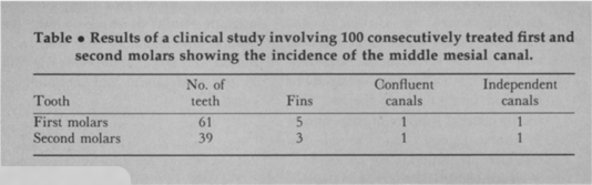
N= 100 1st (61) and 2nd (39) mandibular molars that were consecutively treated in a private endodontic practice
Materials/Methods:
- Fin, the instrument could pass freely between the MB or ML canal and the middle mesial canal.
- Confluent when the prepared canal originated as a separate orifice but apically joined the MB or ML canal.
- Independent when the prepared canal originated as a separate orifice and terminated as a separate foramen. a broad single mesial canal in which three master cones could be cemented to the apex
- After complete serial preparation of other canals, if a catch or possible orifice for MM canal was found, the canal was negotiated.
Most highlighted Results:
Out of the 100, a middle mesial canal was identified and instrumented in 12 cases. (7 in 1st molar, 5 in 2nd molar).
Clinical significance:
It is most important to locate, debride and obturate the middle mesial canal during routine RCT of lower molars.