
Summary:
Purpose: To undertake a comprehensive literature review of the root and root canal morphology of the mandibular first premolar.
Materials/Methods:
Search topics included “number of roots,” “number of canals,” “root canal morphology,” “extra roots,” “anomalies,” and “abnormal morphology.”
- Studies of the mandibular first premolar identified through PubMed plus hand searching
- Over 6,700 permanent mandibular first premolars were analyzed in the studies contained in this review
- Number of roots 2. Number of canals and apical foramina 3. Ethnic differences 4. Sex differences 5. Summary of case reports of other anomalies
Most highlighted Results:
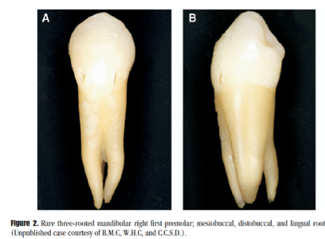
-# of roots > 8 anatomical studies: 98% were single-rooted, 2 roots was 1.8%, 3 roots in 0.2% of the teeth studied.
-Internal canal morphology> 16 anatomical studies: single canal 75.8% of the teeth, Two or more canals found in 24.2% of the teeth.
-10 anatomical studies examined the apical anatomy: One apical foramen was found in 78.9% of the teeth and 21.1% had two or more apical foramina.
Clinical significance:
The root and root canal morphology of this tooth can be complex and requires careful evaluation prior to root canal therapy.