
Purpose: To investigate the morphology of the root canals of extracted human mandibular 1st molar.
N= 45 extracted mandibular 1st molars from 17-22 years old patients.
Materials/Methods:
- Teeth were accessed and root canals were debride
- Teeth placed in a 3% solution of H2O2 for 3 weeks.
- Polyester casting resin stained with a red pigment was placed in the pulp chamber of each tooth to make plastic casts of the root canals.
- Teeth were decalcified in a 35% nitric acid for 10 days.
Most highlighted Results:
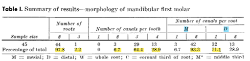
Clinical significance:
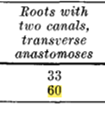
Dentists must always look for a 2nd canal in the distal root of a mandibular first molar. The knowledge that high percentage of teeth contains transverse anastomoses is essential for adequate irrigation activation