Summary:
- Purpose: investigate the anatomic features of the pulp floor and root canal system in 3 rooted mandibular 1st molars.
- n= 122 mandibular 1st molars Inclusion criteria: native Chinese, intact roots.
- n= 25 (2 rooted), 20 (3 rooted) mandibular 1st molars.
Materials/Methods:
- To calculate the frequency, each extracted tooth was stored in 10% formalin and its age, sex, side, and root number of the specimen were recorded.
- To study the morphology of the pulp floor. Teeth were cleaned with 5% NaOCl and ultrasonic scaler. Then scanned at fixed resolution using micro CT. Opacity, view angle and magnification were adjusted to study the root canal system.
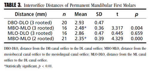
- Interorifice distance measured between all orifices.
- Angle formed by DBO-DLO and MLO-DLO was measured (fig1).
- Vertucci classification was used.
Most highlighted Results:
- frequency of three-rooted teeth was 31.97%.
- 3 rooted teeth had mostly 4 canals, 2 rooted teeth had mostly 3 canals. The third root usually curved severely in proximal view.
- Grooves were often present between DBO and DLO in the three-rooted first molars.
- significantly longer interorifice distance in distal orifices than mesial orifices 2.93 as shown in Table.3.
- Mean angle formed by DBO-DLO and MLO-DLO was 75⁰±10 ⁰.
- In both 3 and 2 rooted 1st molars: mesial roots had type II (2-1), while distal canals had type I (1-1)
- DL root rarely has lateral canals, most of the lateral were found in mesial root of 2 rooted mandibular molar.
Clinical significance:
Understanding the anatomy of 3 rooted mandibular 1st molar would aid in both access and instrumentation.
**(DBO:distobuccalorifice, DLO:distolingualorifice, MLO:mesiolongual orifice, MR: mesial root)
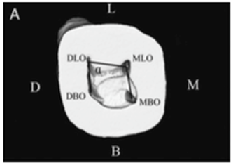
Figure.1