Root Canal Configuration of the Mandibular First Premolar
Authors: Baisden MK, Kulild JC and Weller RN
Year: 1992
Journal: JOE
Summary:
Purpose: To describe the internal anatomy of the mandibular first premolar, the shape of the root canal system, the type of canal configuration, the level at which multiple canals bifurcate, and other variant anatomy were determined.
N = 106 mandibular 1st premolars.
Materials and methods:
- Extraction was due to caries, trauma, periodontal disease or orthodontic reasons.
- A groove was made on the facial side of all teeth which extended from CEJ to the apex at a 3-mm intervals and all sections were examined using stereomicroscope and photographed.
- The classification used to describe the canals was Weine classification
- Additionally, the incidence of multiple canals, the level of canal bifurcation, and any other variant anatomy were noted.
Most highlighted results:
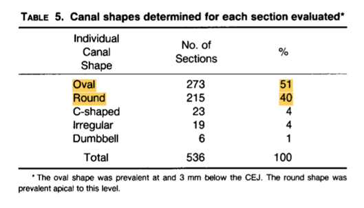
- The shape of root canal was predominantly oval or round.
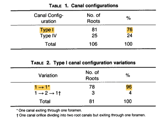
- 76% had type I canal configuration ( which had 96% 1 – 1 )
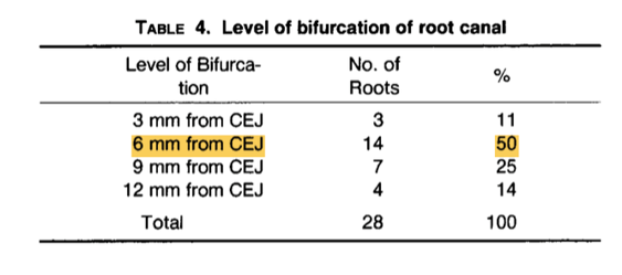
- In 50% of the cases, the bifurcation was found 6 mm from CEJ
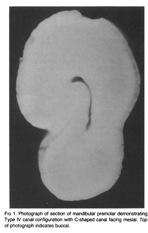
- The number of C- shaped canals occurred in 14% of the roots. (which were associated with Type IV canal systems)
Clinical Significance:
A thorough knowledge of these morphological root canal complexities will enable the clinician to better clean and shape the root canal system.


