Root Canal Anatomy of the human Permanent Teeth
Authors: Vertucci F, Gainesville F.
Year: 1984
Journal: Oral Surgery, Oral Medicine, Oral Pathology journal
Summary:
- Purpose: to do a detailed investigation about root canal anatomy.
- N= 2400 permanent teeth.
Materials/Methods:
- Immediately after extraction, teeth were fixed with 10% formalin.
- Pulp was dyed by injecting hematoxylin dye using 25 gauge needle.
- Teeth were cleared.
- Samples were viewed using dissecting microscope.
Most highlighted Results:
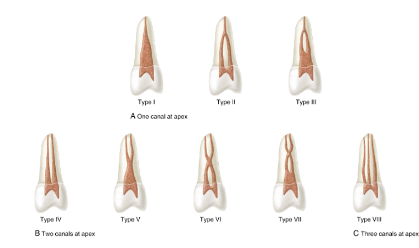
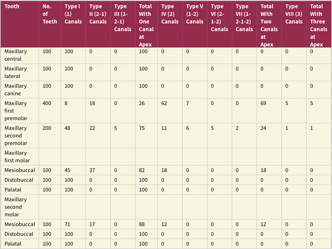
- Upper anteriors have 100% type I.
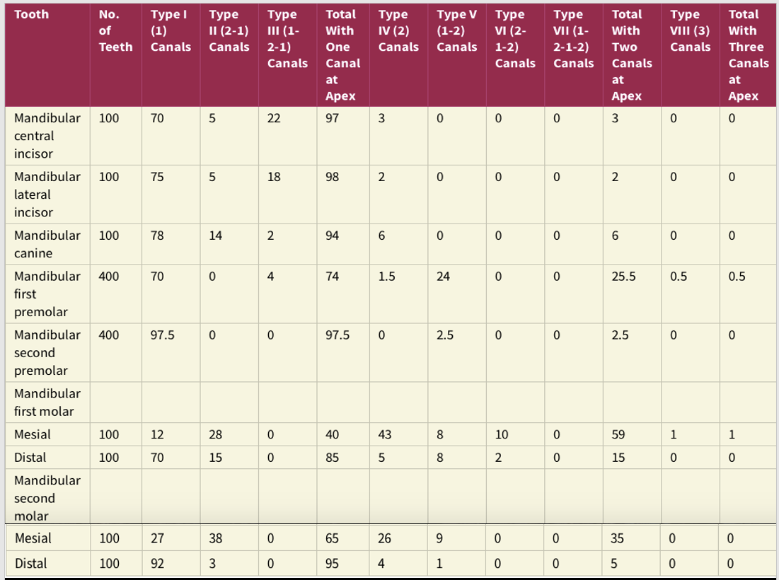
- Mandibular anteriors are 70-80% type I.
- MB2 is more common in MAX. 1st molar (37%) than MAX. 2nd molar (17%). While extra distal is comparable for MAN. 1st (43%), 2nd (38%). Regardless of canals configuration apically.
- 24% of the cases MAN. 1st premolar is type V. MAX. 2nd premolar 11% type IV.
- In all roots you would mostly find 1 foramen, except for MAX. 1st premolar (69%) and mesial root of MAN. 1st molar (.
- Lateral canals are more common in the apical 3rd in all teeth.
- Lateral canals at the furcation is 11% in MAX. 1st premolar and 23% in MAN. 1st molar.
Clinical significance:
- Knowing the diversity/complexity of root canal system would aid clinician predicting , diagnosing & providing higher quality treatment.


