Summary:
Purpose: to identify, treat, and record the number of canals and their relationship to each other in the maxillary molars of the human dentition.
N= Maxillary 1st molar : 1096, max. 2nd : 611 , max .3rd : 25
Materials/Methods:
- Data was obtained from patients’ records treated from 1989 to December 1997.
- The MB2 canal was considered to be present if the author was able to instrument the canal to a depth of 3 to 4 mm after a troughing process.
Most Highlighted Results:
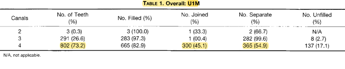
- Over all result : For max 1st molar : MB2 canals were present in 802 (73.2%)
- For max 2nd molar: MB2 canals were present in 310 (50.7%)
- For max 3rd molar: terminus. MB2 canals were only present in 5 (20.0%) teeth
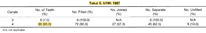
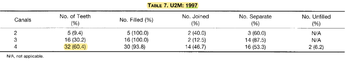
- Results obtained in last 2 years of the study 1996, 1997 (which they used microscope):
- MB2 canals were located in 93.0% of first molars and 60.% in second molars.
Clinical Significance
- When treating Max. molars , using DOM is important in order to detect MB2 canal

After a dental operating microscope (DOM)