
Summary:
Purpose: to determine MB root canal configuration and incidence of MB2 in maxillary 1st molar.
N=208 extracted maxillary 1st molars.
Materials/Methods:
- from the mesial side of the tooth, teeth were sectioned in buccolingual direction using coarse sandpaper disk.
- Then they were viewed and classified.
Most highlighted Results:
- 3 canals configuration were observed:
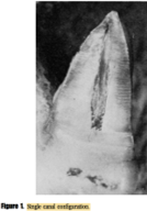
- Type I: single canal/single foramen.(48.5%).
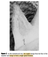
- Type II: 2 canals/ 1 foramen, joining 1-4 mm from apex.(37.5%).
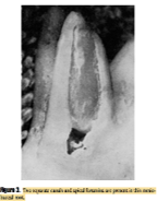
- Type III: 2 canals/2 foramina.(14%).
- Type IV: 1 canal / 2 foramina
Clinical significance:
- Established simple classification system (aid in communication).