Summary:
Purpose:
To investigate anatomical land mark of apical area related to maxillary anterior teeth
N= (30 Max central incisors,30 Max lateral incisors, 30 Max cuspids)
Materials/Methods:
- The root of the tooth cut perpendicularly at the neck, then sequentially ground apex in steps of 50 μm, after that stained with 0.1% violet solution
- Then observed under a stereomicroscope and photographed
Most highlighted Results:
- The root apex and apical foramen at the central incisors and the cuspids were displaced distolabially; while lateral incisors were displaced distolingually.
- 16.7% coincidence between root apex and apical foramen in central incisors and cuspids, while 6.7% of lateral incisors.
- The apical foramen center: displaced mesiolabially in central incisors and the cuspids , and distolingually in lateral incisors.
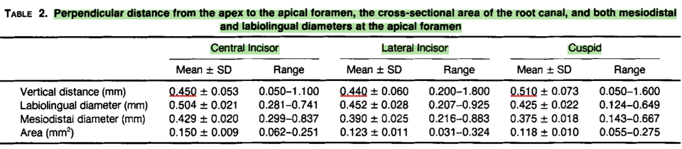
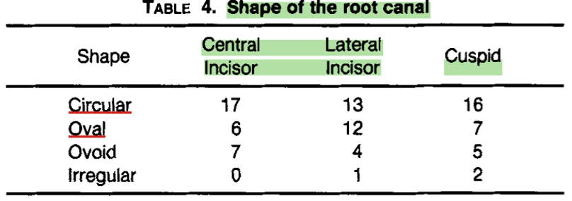
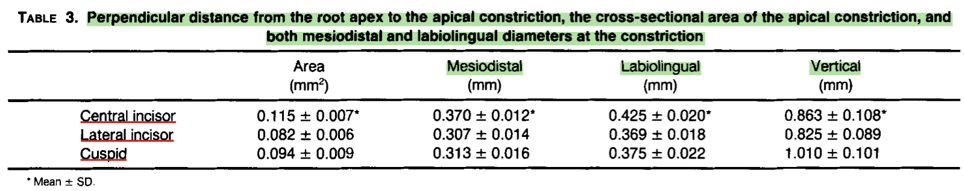
- The rest of parameters measured were in the tables
Clinical significance:
Knowledge about root canal anatomy & morphology is essential to avoid mishaps