Purpose: To assess the variations in the canal system of the palatal roots of maxillary molars.
Summary:
- Not enough studies concerning variances of the root canal system in the palatal roots of maxillary molars, Thews et al in 1979 have reported two clinical cases of aberrant anatomy of the palatal roots of maxillary first molars
- During the pre-clinical course at the University of Illinois, approximately 500 extracted molars were examined in one year.
- Although the incidence was less than 2% it is important to pay attention to this variation.
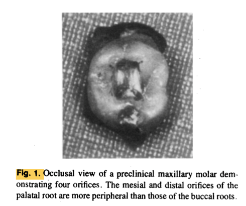
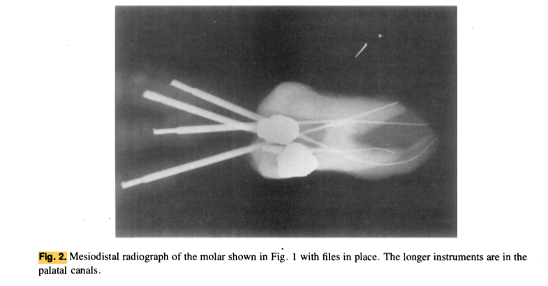
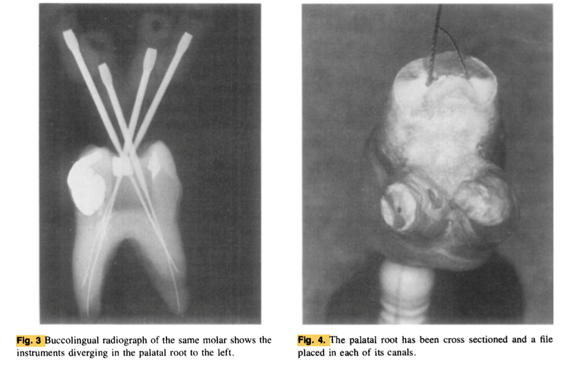
- Figures 1 – 4 show the preclinical cases reported.
- Generally when two canals do exist, more often than not they join together near the apex to form a single canal with a major single communication to the periapex, which certainly will lend itself to successful obturation, even if the dentist fails to find the second canal.
- Moreover, Figures 6 – 7 show clinical cases in which the palatal canal showed two canals.
- To detect these variations clinically, radiographic techniques, with multiple angles are used to better demonstrate this three-dimensional space.
- These techniques are helpful in identifying the variables which, in turn, leads to better treatment with, it is to be hoped, greater success.
Clinical Significance:
The practitioner should be aware of variations in the canal system of the palatal roots of maxillary molars so that they can be treated more effectively.