Root and Root Canal Morphology of Maxillary First Premolars: A Literature Review and Clinical Considerations
Authors: Ahmad IA, Alenezi MA
Year: 2016
Journal: J Endod
Summary:
Purpose: To review the available literature with respect to the root and root canal morphology of maxillary first premolars and discuss the clinical considerations of this morphology on the various dental procedures.
Materials and methods:
- MEDLINE/PubMed and Scopus databases were searched for relevant literature.
- The identified publications were classified into anatomic studies and clinical case reports.
- The data extracted from anatomic studies were tabulated, and weighted averages for certain internal and external morphologic features were calculated.
- The anatomic and developmental variations in the clinical case reports were summarized.
- A total of 92 studies (45 anatomic studies and 47 case reports) including a total of 11,299 teeth were identified.
Most highlighted results:
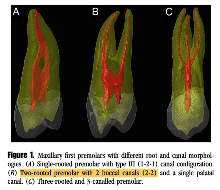
- The majority of maxillary first premolars 56.6% had 2 roots, 41.7% had 1 root, and 1.7% had 3 roots.
- Regardless of the number of roots, the vast majority 86.6% had 2 root canals.
- Type IV (2-2) being the most common canal configuration 64.8%.
- The majority of the apical foramina 66.6% did not coincide with the apical root tip.
- About 38% of the teeth had lateral canals, 12.3% had apical deltas, and 16% had isthmus.
- The clinical case reports showed that the 3-rooted variant was the most common anatomic variation, and developmental anomalies were rarely reported.
Clinical Significance:
Knowledge about the internal and external morphology of maxillary first premolars is important to improve the treatment outcome of surgical and nonsurgical dental procedures.

